How Humans See Color
Many people have seen the below image, often being passed along in friends’ posts. We stare at the image for some time, then look to a white background. We see the opposite image. Green becomes red, yellow becomes blue, and black appears white. The biological reasons for this phenomenon tells us much about human perception of color.
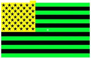
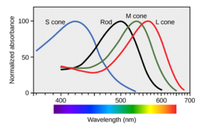
The human eye contains rods (1 type) and cones (3 types) that receive light of certain wavelengths and transmit those signals to the brain. The brain processes the signals into a fully-colored image. The rods process the intensity of light, its lightness. The cones are sensitive to colors: short cones are sensitive to blue, medium cones are sensitive to green, and long cones are sensitive to red wavelengths of light.
So, what happened when we stared at the reverse image of the US flag? When we stare at an object the rods and cones become saturated with information. The afterimage that we see when we stare at a whitespace is a negative inference to the image. Our medium cones are saturated with “green” information, they become tired. When the tired cells look at a white image, they don’t respond as well, and we see the opposing color: red.
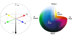
These opposing colors are how humans’ brains process color information. This opponent process theory is the basis for much of color science. Colorists’ models are built upon the biological response of humans.
By the flag example, we can quickly show three opposing channels of visual information. There is a red-green channel; a blue-yellow channel; and a lightness-darkness (white-black) channel. If we plot these channels, we can obtain a three-dimensional sphere. It is these three dimensions that colorists have defined as the LAB color space.
The above discussion makes everything seem logical, except for a glaring omission: we have cones specific for red, blue, and green – but none for yellow. To make yellow, your brain takes some information from the overlapping green and red cones, and some information from the rods and processes it into the color yellow. You don’t see yellow directly, but it is an invented (calculated or processed) color that your brain makes up.
From someone who has managed color matching laboratories, I can attest that yellows (tans and browns) are always the most challenging colors to match. Two highly-trained colorists will view the same yellow to different tolerances. The differences seen are because the two colorists’ brains make up slightly different yellows.
Spectrophotometers can (generally) accurately measure all colors; while humans are not so good at tolerancing. This leads us to a quick refresh on the color tolerances as defined by the CIE in their 1931 specification. The circles shown on the below diagram are MacAdam ellipses (they are 10 MacAdam units). A MacAdam ellipse of one unit show regions where two humans generally say they perceive the same color.
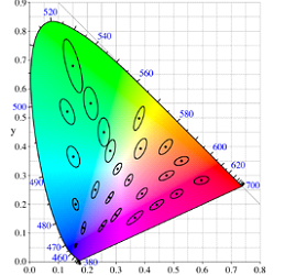
We can see that yellows are highly “squished,” having small ellipses and not much area. This, again, points that two people generally do not tolerate small differences in yellow, like they would for the large areas for green. So when we disagree with someone on the shade of a yellow, let’s agree that we have different brains making it almost impossible to “see” the same color.
To get more technical support, click the picture below!